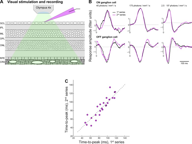
Fig. 7.
Visual sensitivity of the retina-with-choroid in vitro preparation is robust to stimulation at high light intensities. A: horizontal cell and ganglion cell recordings in this study were obtained from the whole mount guinea pig retina with intact retinal pigment epithelium and choroid (GCL, ganglion cell layer; IPL, inner plexiform layer; INL, inner nuclear layer; OPL, outer plexiform layer; ONL, outer nuclear layer; RPE, retinal pigment epithelium; CRD, choroid). Green shading indicates the stimulus light path; magenta shading indicates the glass recording pipette with Alexa 568. B: to test if changes in visual responses observed at high light levels are reversible, we presented the entire stimulus light-level series twice and compared temporal filters computed from responses recorded during the 1st (black) vs. 2nd series (magenta). Panels show overlay of filters at lowest two light levels and at the highest light level for a single example ON (top) and OFF cell (bottom). C: population data comparing filter time to peak at all 8 light levels during the 1st vs. 2nd recorded series for all recorded cells (n = 3). These data indicate that filter changes with increasing light level are reversible and that visual sensitivity is robust against the high light levels used in the experiments.

