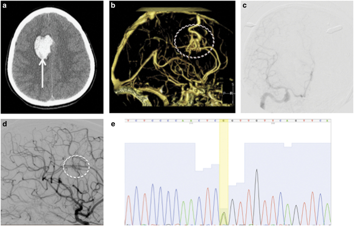
Figure 1.
Clinical features. Initial axial head computed tomography (a) demonstrated a brain hemorrhage (arrow) in the right frontal lobe. Further investigation with computed tomography angiography (b) identified an arteriovenous malformation (circled) as the causative etiology for the hemorrhage. The lesion was resected surgically, and digital subtraction angiography (c) demonstrated no residual arteriovenous shunting. Two years later, a surveillance angiogram was performed (d) that demonstrated regrowth of the arteriovenous malformation (circled). The patient underwent another craniotomy, and the recurrent lesion was removed. Whole-exome sequencing of the tissue was performed, and Sanger sequencing (e) confirmed a mutation at chr13:37439827,G,A (yellow bar) corresponding to a stop codon in SMAD9.

