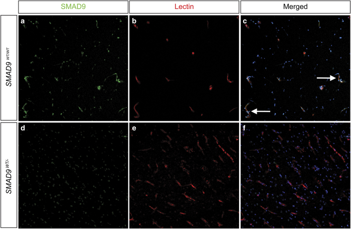
Figure 2.
Effect of the SMAD9 mutation on target protein expression. Immunohistochemical staining was performed to determine the localization and expression of SMAD9 in both control (a, b, c) and lesion (d, e, f) tissue. There was prominent expression of SMAD9 in control tissue (a) that co-localized (arrows) to vascular structures identified by lectin staining of the endothelium (b) and Hoechst nuclear counterstaining (c). Compared to control tissue, SMAD9 expression was greatly reduced in arteriovenous malformation tissue, with the most noticeable loss appearing in perivascular tissue.

