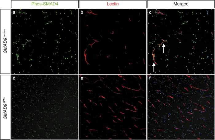
Figure 3.
Effect of the SMAD9 mutation on downstream pathway protein phosphorylated SMAD4. Immunohistochemical staining was performed to determine the localization and expression of p-SMAD4 in both control (a, b, c) and lesion (d, e, f) tissue. Similar to SMAD9 staining, there was prominent expression of p-SMAD4 in control tissue (a) that co-localized (arrows) to vascular structures identified by lectin staining of the endothelium (b) and Hoechst nuclear counterstaining (c). Decreased expression of p-SMAD4 was observed in arteriovenous malformation tissue, and its expression appeared to be dissociated from vascular structures.

