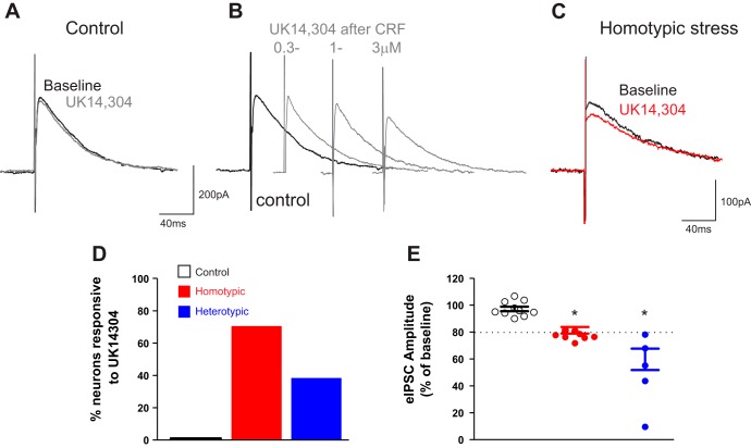
Fig. 4.
Stress and corticotropin-releasing factor (CRF) uncover the α2-adrenoceptor-mediated decrease of evoked inhibitory postsynaptic current (eIPSC) amplitude. A: representative traces of eIPSCs recorded in a dorsal motor nucleus of the vagus (DMV) neuron from control rat following electrical stimulation of nucleus tractus solitarius (NTS). Note that perfusion with UK14304 (1 µM) did not affect the amplitude of eIPSCs. Holding potential = −50 mV. B: representative traces of eIPSCs recorded in a DMV neuron from a control rat. Note that following perfusion with CRF and recovery to baseline amplitude, a second perfusion with UK14304 reduced the eIPSC amplitude in a concentration-dependent manner. Holding potential = −50 mV. C: representative traces of eIPSCs recorded in a DMV neuron from a chronic homotypic stress (CHo) rat. Perfusion of the slice with UK14304 reduced eIPSC amplitude even without pretreatment of the slice with CRF. Holding potential = −50 mV. D: graphic representation of the percentage of neurons in control (solid bar; N = 0/10), CHo stress (red bar; N = 12/17), or chronic heterotypic stress (CHe) stress (blue bar; N = 5/13) in which perfusion with UK14304 decreased eIPSC amplitude. E: graphic representation of the UK14304-induced modulation of the eIPSC amplitude in control, CHo, and CHe stress groups. In the CHo and CHe groups, only responsive neurons are depicted. Dashed line represents the “threshold” change to classify the neurons as responsive. Data were analyzed with 1-way ANOVA followed by Tukey’s test. P < 0.05 vs. baseline.

