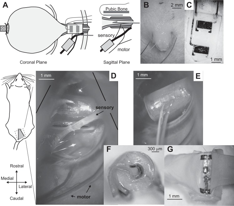
Fig. 1.
Experimental setup. A: placement of the bladder catheter, nerve cuff, and electrodes for recording external urethral sphincter (EUS) electromyogram (EMG). Two different types of EMG electrodes were used (in different experiments), percutaneous wires (B) and flat metal contacts embedded in a silicone substrate (C) that was placed underneath the pubic bone. D: view through the surgical microscope of the isolated sensory pudendal nerve in the ischiorectal fossa. E: view showing placement of the nerve cuff on the sensory pudendal nerve. F: side view of a nerve cuff showing the orientation of the incoming wires, as well as the tabs that are used to open the cuff. G: view of the inside of the nerve cuff after pulling the cuff open at the tabs. The exposed portion of the contacts are near the outside of the cuff.

