Fig. 2.
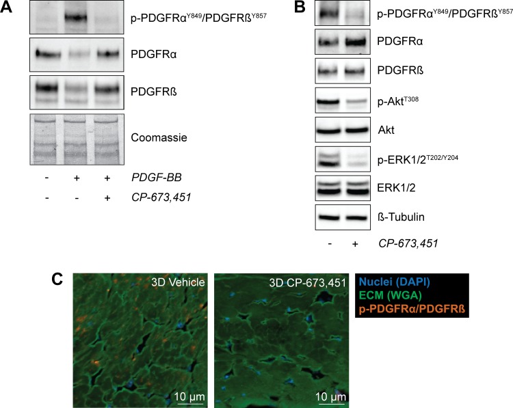
CP-673,451 inhibits the phosphorylation of PDGFRα and PDGFRβ in tendon fibroblasts in vitro and in vivo. A: representative immunoblots of serum-starved tail tendon fibroblasts treated with 20 ng/ml of PDGF-BB for 30 min with or without 1 μM of the PDGFR inhibitor CP-673,451. A Coomassie stained membrane is shown as a loading control. B: representative immunoblots of 3-day overloaded plantaris tendons demonstrating the ability of CP-673,451 to inhibit phosphorylation of PDGFRα and PDGFRβ in vivo. Phosphorylation of Akt and ERK1/2 was also inhibited by PDGFR inhibitor treatment. β-Tubulin is shown as a loading control. C: immunohistochemistry of 3-day overloaded plantaris tendons treated with vehicle or PDGFR inhibitor showing a decrease in the abundance of p-PDGFRα/PDGFRβ-expressing cells in the overloaded plantaris tendons treated with CP-673,451 relative to vehicle-treated controls. Scale bars are 10 μm. DAPI, blue; WGA, green; p-PDGFRαY849/PDGFRβY857, red.

