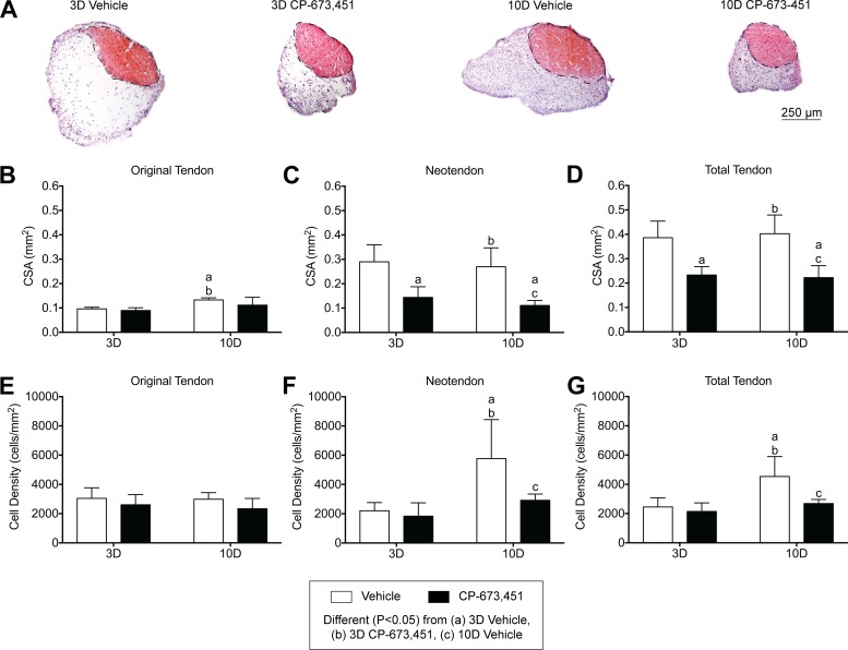
Fig. 3.
Inhibition of PDGFR signaling prevents growth of plantaris tendons subjected to mechanical overload. A: representative cross sections of 3- and 10-day overloaded plantaris tendons, treated with vehicle or PDGFR inhibitor, and stained with hematoxylin and eosin. Dashed black line indicates the boundary between the original tendon and neotendon. Scale bar is 250 μm. B–G: quantitative analysis of cross-sectional area (CSA, in mm2) (B–D) and cell density (cells/mm2) (E–G) for the original tendon, neotendon, and total tendon. Values are mean ± SD; n = 5 tendons for each group. Differences between groups were tested using a two-way ANOVA (α = 0.05) followed by Tukey’s post hoc sorting: different (P < 0.05) from a, 3D vehicle; b, 3D PDGFR inhibitor; c, 10D vehicle.

