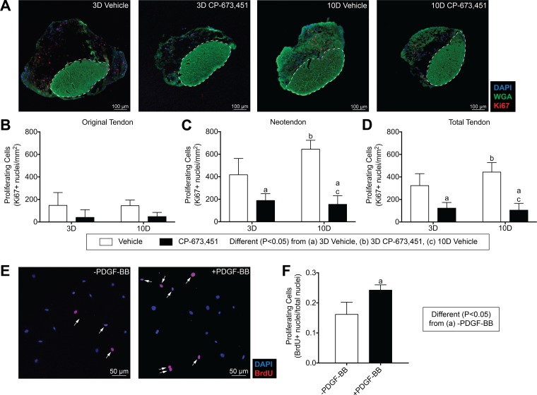
Fig. 4.
Inhibition of PDGFR signaling decreases tendon fibroblast proliferation. A: representative Ki67 immunostaining of 3- and 10-day overloaded plantaris tendons, treated with vehicle or PDGFR inhibitor. Dashed white line indicates the boundary between the original tendon and neotendon. Scale bars are 100 μm. DAPI, blue; WGA, green; Ki67, red. B–D: quantitative analysis of proliferating cells (Ki67+ nuclei/mm2) for the original tendon, neotendon, and total tendon. Values are means ± SD; n ≥ 4 tendons for each group. Differences between groups were tested using a two-way ANOVA (α = 0.05) followed by Tukey’s post hoc sorting: different (P < 0.05) from a, 3D vehicle; b, 3D PDGFR inhibitor; c, 10D vehicle. E: in vitro proliferative activity of tail tendon fibroblasts was measured by BrdU uptake in the presence of 0.5% FBS with or without 20 ng/ml of PDGF-BB treatment. Proliferating cells were double-labeled for DAPI (blue) and BrdU (red) and are marked by white arrows. F: proliferative activity was quantified as means ± SD in 5 randomly selected fields of a single experiment from 6 independent experiments performed. Differences between groups were tested using an unpaired t-test; a, P < 0.05.

