Fig. 10.
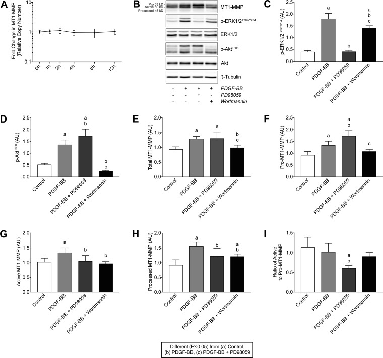
PDGF-BB does not regulate MT1-MMP mRNA expression but does increase MT1-MMP protein levels through a PI3K/Akt-dependent mechanism. A: quantification of MT1-MMP transcript levels in tail tendon fibroblasts treated with 20 ng/ml of PDGF-BB for 1, 2, 4, 8, and 12 h. Values are means ± SD; n ≥ 3 replicates. Differences between groups were tested using one-way ANOVA (P < 0.05). B: representative immunoblots of Pro-MT1-MMP (63 kDa), active MT1-MMP (60 kDa), processed MT1-MMP (45 kDa), and phospho and total ERK1/2 and Akt are shown, from tendon fibroblasts incubated alone or with PDGF-BB in the presence or absence of PD98059 or wortmannin for 24 h. β-Tubulin is shown as a loading control. C–H: quantification of p-ERK1/2T202/Y204 (C), p-AktT308 (D), total MT1-MMP (E), pro-MT1-MMP (F), active MT1-MMP (G) and processed MT1-MMP (H) protein levels. AU, arbitrary units. I: ratio of the pro- to active form of MT1-MMP. Values are means ± SD; n = 6 replicates. Differences between groups were tested using a one-way ANOVA (α = 0.05) followed by Tukey’s post hoc sorting: different (P < 0.05) from a, control; b, PDGF-BB; c, PDGF-BB + PD98059.

