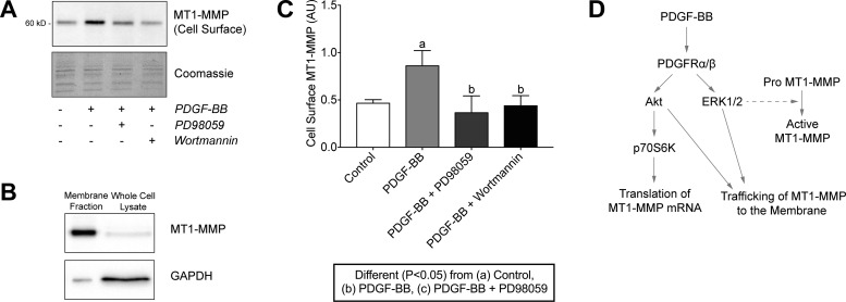
Fig. 11.
PDGF-BB-dependent membrane trafficking of MT1-MMP in tendon fibroblasts is regulated by the PI3K/Akt and ERK1/2 pathways. (A) Representative immunoblots of cell surface MT1-MMP protein levels in tail tendon fibroblasts after incubation alone or with PDGF-BB in the presence or absence of PD98059 or wortmannin for 24 h in low-serum conditions. Coomassie staining is shown as a total protein loading control. (B) Representative immunoblots of the membrane fraction and whole cell lysates of tendon fibroblasts using MT1-MMP and GAPDH as typical membrane and cytosolic proteins, respectively. Due to the isolation detergents, membrane fraction lanes run in a narrow fashion. (C) Quantification of cell surface MT1-MMP protein levels. Values are means ± SD; n ≥ 3 replicates. Differences between groups were tested using a one-way ANOVA (α = 0.05) followed by Tukey’s post hoc sorting: different (P < 0.05) from a, control; b, PDGF-BB; c, PDGF-BB + PD98059. D: diagram of PDGF-BB-dependent regulation of MT1-MMP expression and membrane trafficking in tendon fibroblasts.

