Fig. 2.
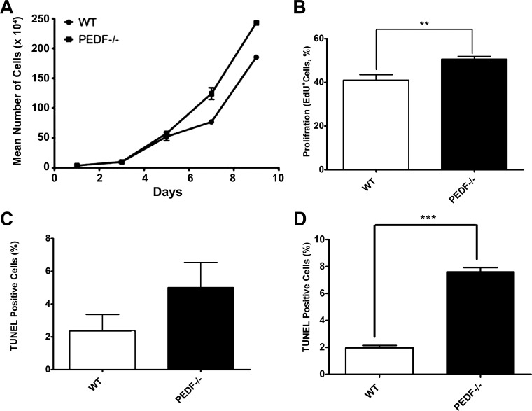
Alterations in proliferation and apoptosis of ChECs. PEDF deficiency resulted in decreased proliferation of ChECs. A: the rate of cell proliferation was determined by counting the number of cells. A significant increase in the proliferation of PEDF−/− ChECs was observed compared with PEDF +/+ cells. B: an increase in the rate of DNA synthesis was observed in PEDF−/− ChECs compared with the PEDF+/+ cells. C: the rate of apoptosis was determined using TdT-dUTP terminal nick-end labeling (TUNEL). A similar experiment was carried out using H2O2 (200 μΜ) as an inducer of apoptosis (positive control). No significant differences were observed in the basal rate of apoptosis in these cells. D: a significant increase in the rate of apoptosis was observed in PEDF−/− ChECs when challenged with H2O2 (200 μΜ) compared with PEDF+/+ cells (**P < 0.01, ***P < 0.001, n = 3).

