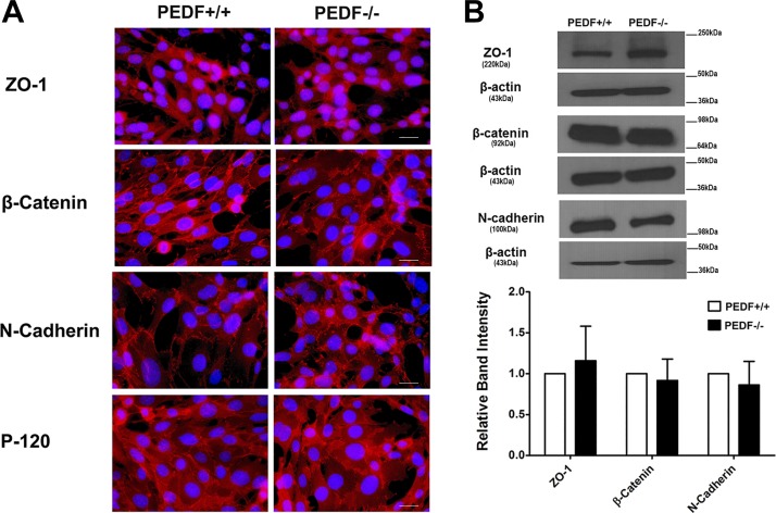
Fig. 3.
Cellular localization and expression of junctional proteins. A: the localization of ZO-1, N-cadherin, β-catenin, and P-120 catenin was determined by immunofluorescence staining. The PEDF+/+ and PEDF−/− ChECs were plated on fibronectin-coated chamber slides and stained with specific antibodies as detailed in materials and methods. No staining was observed in the absence of primary antibody (not shown). No significant difference was observed in localization of these proteins (scale bar = 20 µm). B: Western blot analysis of junctional proteins. Total cell lysates were prepared from PEDF+/+ and PEDF−/− ChECs and analyzed for expression of ZO-1, N-cadherin, β-catenin, P-120 catenin, and β-actin. β-Actin was used for loading control. The quantification of data is shown in the panel at bottom. No significant differences were observed in the levels of these proteins. These experiments were repeated using two different isolations of ChECs with similar results (n ≥ 3).

