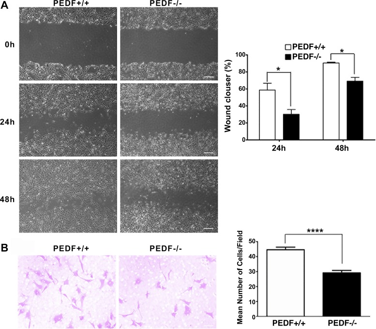
Fig. 4.
Attenuation of migration in PEDF−/− ChECs. A: scratch wound assay of ChEC monolayers on gelatin-coated plates was used to determine cell migration. Wound closure was observed by phase microscopy at indicated time points (scale bar = 100 µm). The quantitative assessment of wound migration is shown on the right (*P < 0.05; n = 3). B: migration of ChECs in transwell assays. Note the significant decrease in the migration of PEDF−/− ChECs compared with PEDF+/+ cells (*P < 0.05, ****P < 0.0001, n = 6; scale bar = 20 µm).

