Fig. 7.
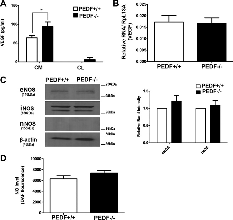
Alterations in VEGF and NOS expression. A: the level of VEGF was determined in conditioned medium (CM) or cell lysates (CL) collected from PEDF+/+ and PEDF−/− ChECs using an ELISA as detailed in materials and methods. Note the significant increase in the level of VEGF (CM) produced by PEDF−/− ChECs compared with PEDF+/+ cells (*P < 0.05, n = 3). B: the level of VEGF mRNA was determined by qPCR using RNA prepared from ChECs. There was no significant difference in the amount of VEGF mRNA detected in ChECs (P > 0.05, n = 3). C: the levels of eNOS, iNOS, and nNOS were determined by Western blot analysis of cell lysates. There was no significant difference in the level of eNOS and iNOS in ChECs. We did not detect nNOS expression in ChECs. β-Actin was used as a loading control. D: the level of intracellular NO was determined using DAF-FM as detailed in materials and methods. Note similar NO levels in all cells (P > 0.05, n = 3).

