Figure 1.
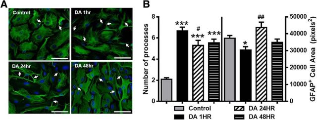
Elevated extracellular DA induces morphological changes in astrocytes. A, Widefield images of astrocytes double-stained for GFAP (green) and DAPI (blue) in control conditions and after exposure to DA (75 μm) for 1, 24, or 48 h (DA 1 h, DA 24 h, DA 48 h). DA induces a prominent increase in the number of primary processes (arrows). Scale bars, 75 μm. B, Quantification of the number of primary processes and average GFAP+ cell area. One-way ANOVA for number of processes (F(3,469) = 56.64, p < 0.0001). One-way ANOVA for GFAP+ area (F(3,469) = 5.52, p = 0.001). Bonferroni post hoc: *p < 0.05 versus control. ***p < 0.001 versus control. #p < 0.05 versus DA 1 h. ##p < 0.01 versus DA 1 h. Control, n = 179 cells; DA 1 h, n = 152 cells; DA 24 h, n = 58 cells; DA 48 h, n = 84 cells.

