Fig. 2.
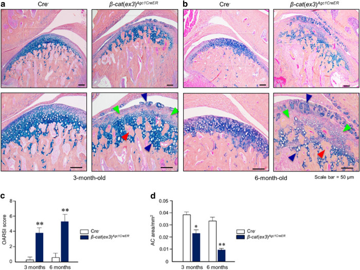
β-Catenin conditional activation mice (β-cat(ex3)Agc1CreER) show a progressive osteoarthritis (OA)-like phenotype in temporomandibular joint (TMJ) tissue. a TMJ samples were dissected from 3- and 6-month-old mice and Alcian blue/hematoxylin staining was performed. β-cat(ex3)Agc1CreER mice displayed early signs of an OA-like phenotype, including increased numbers of hypertrophic chondrocytes, the rough articular surface (blue arrowheads), loss of articular chondrocytes (green arrowheads), and new woven bone formation inside hypertrophic chondrocyte areas (red arrowheads). b Loss of cartilage tissue (green arrowheads), increased numbers of hypertrophic chondrocytes and rough articular surface (blue arrowhead), and new woven bone formation inside the hypertrophic chondrocyte areas (red arrowhead) were observed in 6-month-old β-cat(ex3)Agc1CreER mice. c Analysis using the scoring system recommended by the Osteoarthritis Research Society International (OARSI) revealed cartilage destruction in 3- and 6-month-old β-cat(ex3)Agc1CreER mice (**P < 0.01, versus Cre− mice; n = 5 per group). d Histomorphometric analysis showed that TMJ cartilage areas were significantly reduced in 3- and 6-month-old β-cat(ex3)Agc1CreER mice (*P < 0.05, **P < 0.01, versus Cre− mice; values are expressed as mean ± standard errors; n = 5 per group)

