Fig. 5.
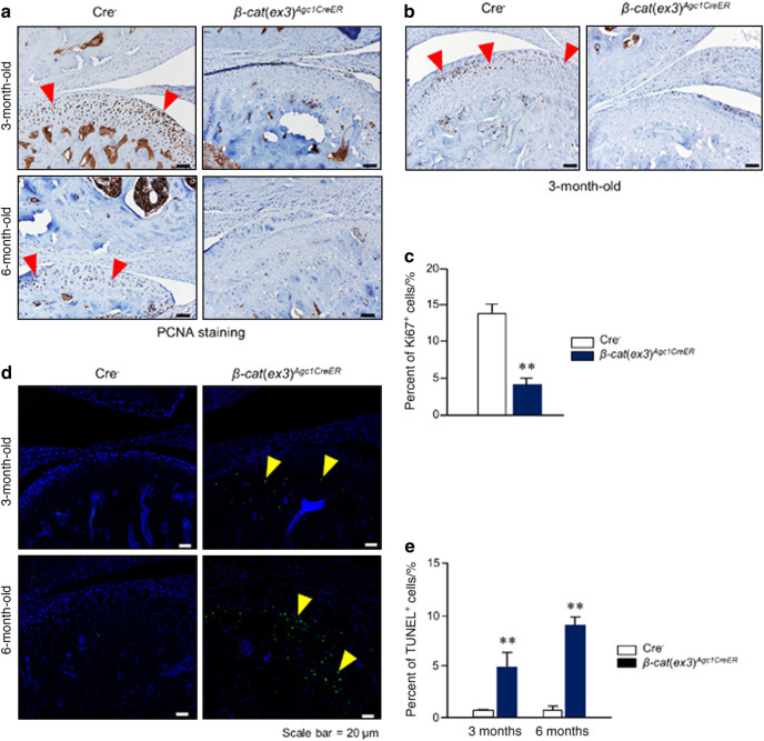
Alteration of cell proliferation and apoptosis in 3- and 6-month-old β-cat(ex3)Agc1CreER conditional activation mice. a Results of proliferating cell nuclear antigen (PCNA) staining revealed that cell proliferation was significantly reduced in temporomandibular joint (TMJ) chondrocytes in β-cat(ex3)Agc1CreER mice. Red arrowheads: PCNA-positive cells. b, c Results of Ki67 staining revealed that cell proliferation, mostly in the middle layer of condylar chondrocytes, significantly reduced in TMJ cartilage in 3-month-old β-cat(ex3)Agc1CreER mice. Red arrowheads: Ki67-positive cells. d, e Results of terminal deoxinucleotidyl transferasemediated dUTP-fluorescein nick end labeling (TUNEL) staining demonstrated increased numbers of apoptotic cells were detected in TMJ cartilage of β-cat(ex3)Agc1CreER mice (**P < 0.01 versus Cre− mice; values are expressed as mean ± standard errors; n = 5 per group). Yellow arrowheads: TUNEL positive cells

