
Figure 4. G-CSF Increases Total Microglial Activity Selectively in Tg APP+PS1 Mice.
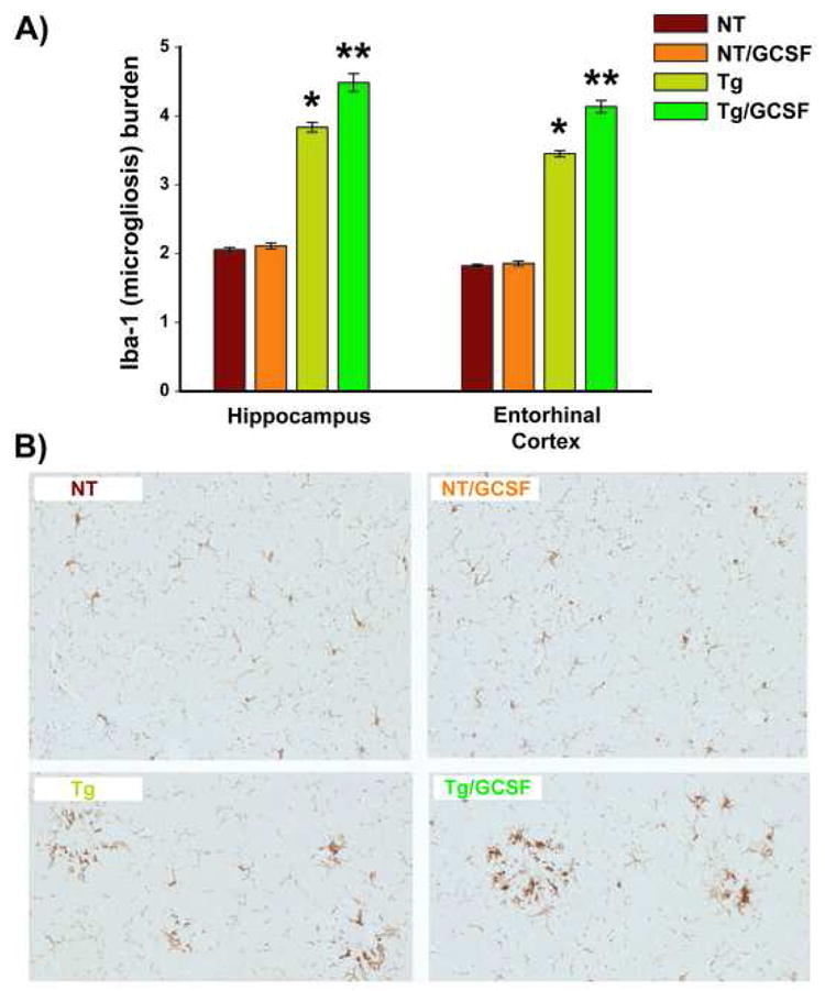
(A) Increased microgliosis was evident by quantitative Iba1 immunostaining in both hippocampus and entorhinal cortex of Tg/GCSF mice, but not NT/GCSF mice; * p < 0.00001 versus both NT groups, **p<0.00001 versus Tg group. (B) Saline-treated Tg APP+PS1 mice exhibited clusters of microglia surrounding amyloid, while G-CSF treated Tg mice revealed a significant increase in Iba1 immunoreactivity around and within amyloid deposits in hippocampus (B) and entorhinal cortex (not shown). Scale bar = 50 μm.