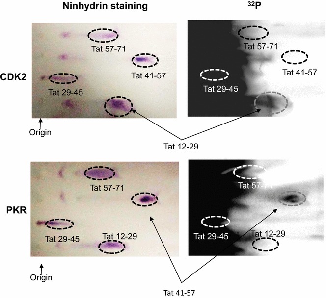
Fig. 3.

Hunter peptide mapping analysis of Tat phosphorylation by CDK2 and PKR. Tat-derived peptides were phosphorylated in vitro by CDK2/cyclin E or PKR, as indicated. The reactions were loaded on nitrocellulose plates and peptides were resolved by thin layer electrophoresis as described in Methods. Plates were dried and stained with ninhydrin (left panels) or exposed to Phospho Imager screen (right panels). Origin and peptide positions are indicated on figure. The results are representative from 2 experiments
