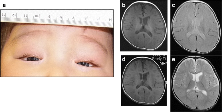
Fig. 1.

Clinical features of the patient. a The face at 5 months of age. The patient had blue irides, blepharoptosis, and dystopia canthorum (W-index: 2.24). b-e Brain MRI findings at 8 and 18 months of age. At 8 months of age, namely, 3 months after the proband had a 4-min-long systemic clonic seizure, T1- b and T2-weighted images c demonstrated delayed myelination of the frontal lobe. In normal development, myelination of frontal lobe is observed at 3–4 months of age on T1-weighted imaging, and at 7–8 months of age on T2-weighted imaging. Follow-up MRI performed at 18 months of age demonstrated almost normal myelination on T1-weighted imaging d and T2-weighted imaging e
