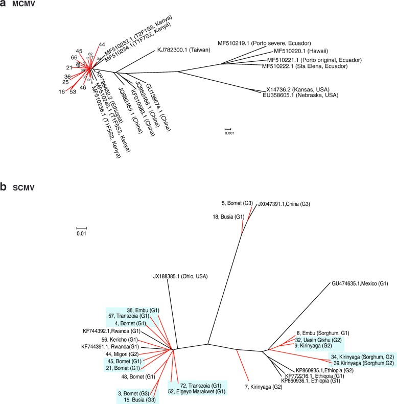
Fig. 7.
Phylogeny of MCMV (a) and SCMV (b). Phylogenetic trees were generated using Bayesian inference in Mr. Bayes 3.2. Scale bar represents nucleotide substitution per site. For SCMV, G1, G2 and G3 correspond to genetic variation and groups described in Fig. 4. Kenya samples described in this study are colored in red and identified by a number and the county of origin. Unless indicated otherwise, samples came from maize. Green background indicates clusters formed by Kenya samples

