Fig. 3.
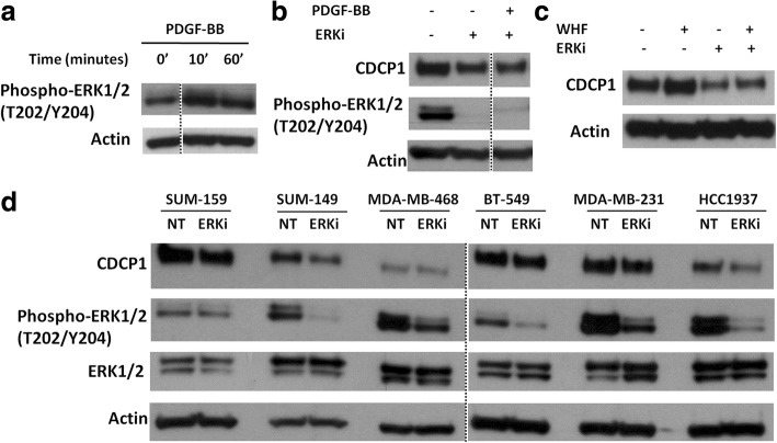
ERK1/2 activity regulates CDCP1 expression in TNBC cells. a WB analysis of phospho-ERK1/2 (T202/Y204) in MDA-MB-231 cells treated with or without 20 ng/ml PDGF-BB for 10 and 60 min. b WB analysis of CDCP1 and phosphoERK1/2 (T202/Y204) in MDA-MB-231 cells treated with or without the ERK1/2 inhibitor UO126 (2 μM) and stimulated with or without 20 ng/ml PDGF-BB for 24 h. c WB analysis of CDCP1 in MDA-MB 231 cells treated with or without UO126 (2 μM) and stimulated with or without 5% WHF in culture medium for 24 h. Dotted lines demarcate juxtaposed images originating from separate lines of the same western blot. d WB analysis of CDCP1, phosphoERK1/2 (T202/Y204), and ERK1/2 in SUM149, SUM159, MDA-MB468, BT-549, MDA-MB-231, and HCC1937 cells treated with or without UO126 (2 μM) under standard medium conditions for 24 h. Monoclonal anti-actin was used as the total protein loading control

