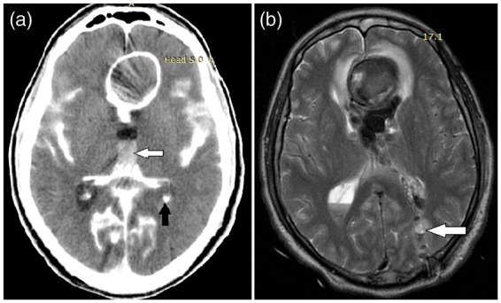
Figure 3.

Computed tomography (CT) scan (a) reveals massive subarachnoid hemorrhage (SAH) with interhemispheric hematoma formation (white arrow) and ventricular breakthrough (black arrow). Postoperative magnetic resonance imaging (MRI) (b) shows position of the external ventricular drainage (EVD) (white arrow).
