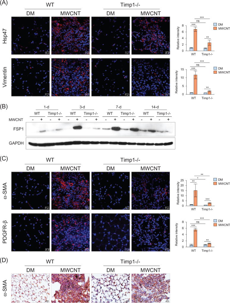
Figure 3.
Recruitment of fibroblasts and myofibroblasts by MWCNTs. Lung sections from mice exposed to DM or 40 µg MWCNTs for 7 d were examined for the expression of fibroblast and myofibroblast markers (A, C and D). (A) Immunofluorescence detection of fibroblast markers Hsp47 and Vimentin. (B) Immunoblotting of FSP1. Lung proteins from randomly selected samples of each group exposed to DM or 40 µg MWCNTs were analyzed and a representative blotting image is presented. (C) Immunofluorescence detection of myofibroblast markers α-SMA and PDGFR-β. (D) Immunohistochemistry staining of α-SMA. Images have scale bars of 20 µm. For (A) and (C), the red color indicates positive staining and blue indicates nuclear staining. Relative intensity is shown as the mean ± SD (n = 4).

