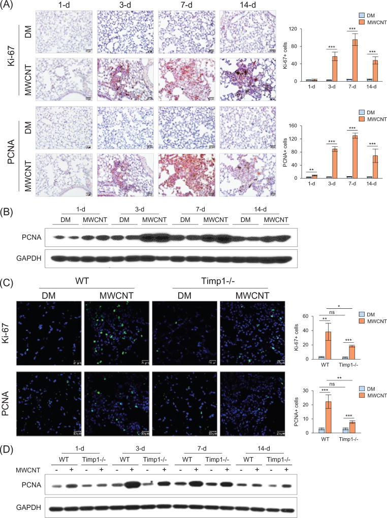
Figure 4.
Reduced cell proliferation in Timp1 KO lungs. (A and B) Time-dependent stimulation of cell proliferation by MWCNTs (40 µg) in WT lungs on days 1, 3, 7 and 14 post-exposure. (A) Immunohistochemistry of Ki-67 (upper panel) and PCNA (lower panel) on lung sections of WT mice. Red indicates positive staining and blue nuclear counterstaining. Scale bar: 20 µm. The number of cells with positive staining is shown as the mean ± SD (n = 4). (B) Immunoblotting of PCNA in lung tissues of WT mice. Lung proteins from two randomly selected samples of each group were used. (C and D) Comparison of cell proliferation between WT and Timp1 KO lungs exposed to MWCNTs (40 µg). (C) Immunofluorescence of Ki-67 (upper panel) and PCNA (lower panel) on lung sections (7 d post-exposure). Green indicates positive staining and blue nuclear staining. Scale bar: 20 µm. The number of cells with positive staining is shown as the mean ± SD (n = 4). (D) Immunoblotting of PCNA. Lung proteins from randomly selected samples of each group were studied, and a representative blotting image is presented.

