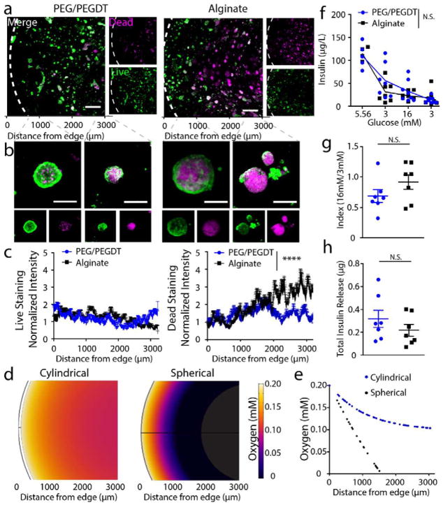
Figure 4. In vitro characterization of encapsulated islet viability and function within 500 μL macrodevices.
Live/dead imaging at (a) low and (b) high magnification demonstrates islet viability in PEG/PEGDT gels compared to alginate. Swelling of PEG/PEGDT post encapsulation results in lower density of islets within macrodevices, reducing detrimental nutrient gradient compared to non-swelling alginate gels and resulting in improved islet viability throughout PEG/PEGDT gels, as quantified in (c) Live and Dead intensity values normalized to edge values. (d) Gel cross section oxygen distribution profiles and (e) quantitative plot of oxygen concentration within gels demonstrate greater nutrient competition in spherical gels compared to cylindrical disk-shaped gels. (f) Glucose stimulated insulin response assay, (g) index values, and (h) total insulin released demonstrate comparable response between alginate and PEG/PEGDT macrodevices. Live/dead histograms evaluated by repeated measures one-way ANOVA with Sidak’s multiple comparison test. GSIR data analyzed by two-way ANOVA with Sidak’s multiple comparison test, n = 7/group, data pooled from two separate experiments. Error bars = SEM, scale bars panel A = 500 μm, scale bars panel B = 50 μm. **** P < 0.0001 by two-way ANOVA, N.S. = no significance.

