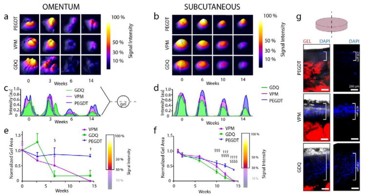
Figure 5. In vivo monitoring of PEG hydrogel macroencapsulation device stability comparing non-degradable synthetic (PEGDT) and degradable peptide (VPM, GDQ) cross-linkers.
RGD adhesive ligand was fluorescently labeled for in vivo tracking of synthetic hydrogels transplanted in the (a, c, e) omentum and (b, d, f, g) subcutaneous space (n = 4/group) of rats. Topographic images of IVIS in vivo gel imaging at select time points demonstrate signal stability and remodeling of gel shape for (a) OM and (b) SUBQ implanted gels. Corresponding signal intensity histograms for cross sections of gels in (a) and (b) illustrate fluorescent signal intensity over time for (c) OM and (d) SUBQ implanted gels. Temporal changes in gel area was quantified from IVIS topographic images (A and B) by thresholding signal at 30% intensity and normalization to early time points for both (e) OM and (f) SUBQ implanted gels. (g) Explanted gels demonstrate greater levels of tissue infiltration for degradable cross-linkers (SUBQ). Gel degradation analyzed by two-way ANOVA with Tukey’s multiple comparison test. Scale bars = 200 μm. $ P < 0.05, $$$ P < 0.0005, $$$$ P < 0.0001 vs. GDQ; † P < 0.05, ††† P < 0.0005, †††† P < 0.0001 vs. VPM.

