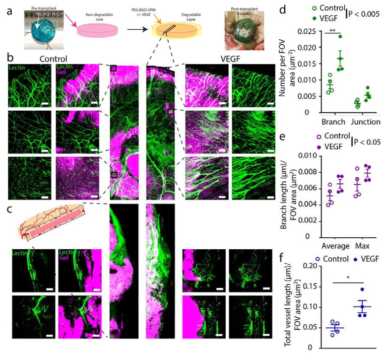
Figure 6. Vascular remodeling of PEG/PEGDT macrodevices transplanted with vasculogenic layer in the omentum.
(a) Fluorescently labeled RGD-laden non-degradable macrodevices were transplanted within the omentum of rats with a degradable (VPM) hydrogel layer, either with (VEGF) or without (Control) vasculogenic factor. At 4 weeks post-transplant, subjects were lectin perfused to label functional vasculature (green), and whole mount imaged to visualize degree of (b) surface and (c) cross section vascularization. Surface vascularization was characterized and quantified for number of (d) vessel junctions and branches, (e) average and maximum branch length, (f) and total overall vessel length per field of view (FOV). (n = 4/condition, FOV = 5–8/n). * P < 0.05, ** P < 0.005. Branch length and branch/junction numbers were analyzed by two-way ANOVA with Sidak’s multiple comparison test. Total vessel length analyzed by Student’s t-test. Scale bar = 200 μm.

