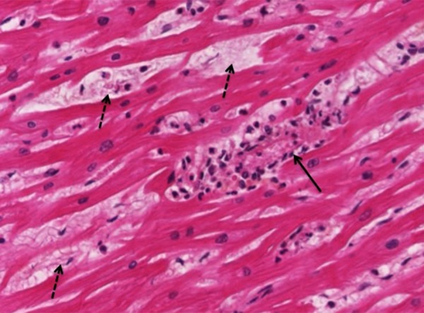
Figure 2.

Illustration of the cardiac tissue, post-mortem histopathological examination (Hemalin Phloxin Safran coloration; 400×). Inflammatory infiltrate with myocardic necrosis is shown in one field of cardiac tissue (solid arrow); multifocal lesions with myocardic edema are shown (dashed arrows).
