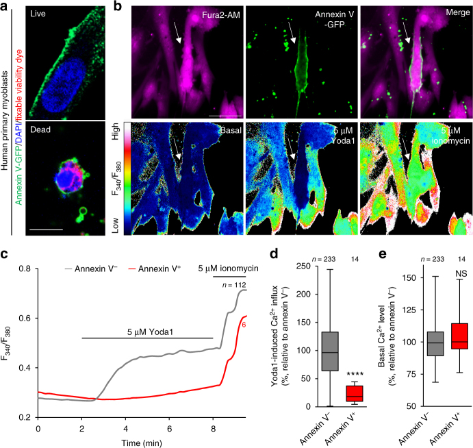
Fig. 4.
Impaired PIEZO1 activation in PS-exposing human primary myoblasts during myotube formation. a Detection of cell surface-exposed PS on differentiating human primary myoblasts by annexin V-GFP (PS, green), DAPI (nuclei, blue) and fixable viability dye (dead cells, red). b Fura2 imaging of Ca2+ influx in annexin V-GFP-labelled human primary myoblasts (arrow) upon addition of Yoda1 and ionomycin. Representative traces (c), quantification of Yoda1-induced Ca2+ influx (d) and basal Ca2+ level (e) in b. ****P < 0.0001 (Student’s t-test). NS not significant, n sample number. Box and whiskers graph―line: median, box: upper and lower quartiles, whiskers: maxima and minima. Scale bars: 10 μm (a), 20 μm (b)

