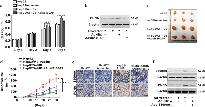
Fig. 5. Involvement of S100A9 in HBx-mediated growth of HepG2 cells.
a CCK8 analysis for HepG2 cells infected with or without Ad-vector, AdHBx, AdHBx and AdsiS100A9 for sequential 4 days. b Western blot analysis for PCNA expression in HepG2 cells infected with or without Ad-vector, AdHBx, AdHBx and AdsiS100A9 for 72 h. c Images of representative mice bearing tumors derived from mice injected with HepG2 cells infected with or without Ad-vector, AdHBx, AdHBx and AdsiS100A9. d Tumor growth curves of all groups. Subcutaneous tumor growth was recorded every 5 days with vernier calipers. e IHC staining analysis for S100A9 and PCNA in representative xenograft tumor sections. Black scale bar = 150 μm. f Western blot analysis for S100A9 and PCNA expression in representative xenograft tumor tissues; *p < 0.05, **p < 0.01

