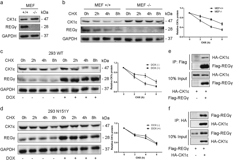
Fig. 4. REGγ interacts with CK1ε and promotes its degradation.
a Expression of CK1ε in REGγ+/+(REGγ wild type) and REGγ−/− (REGγ knockout) MEF cells determined by WB. b REGγ+/+and REGγ−/− MEFs were treated with CHX for the indicated time. The level of CK1ε was detected using WB. Relative quantification of CK1ε levels is shown. c, d CHX chase assay was performed on 293 WT cells with wild-type REGγ (c) and 293 N151Y cells with mutant REGγ (d) following DOX induced overexpression of REGγ. Relative quantification of CK1ε levels is shown. e Interaction between REGγ and CK1ε in 293T cells as determined by coimmunoprecipitation and WB analysis by using Flag beads following transient transfection of Flag-REGγ and HA-CK1ε. f Reciprocal interaction between REGγ and CK1ε as determined by coimmunoprecipitation by using HA beads as indicated. Data are shown as mean ± SD. *P < 0.05

