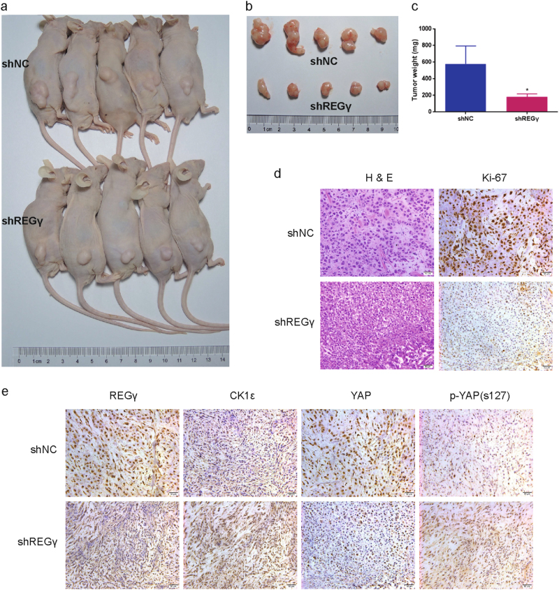
Fig. 7. REGγ depletion suppresses RCC cells growth in vivo.
a ACHN shNC and shREGγ cells were injected into the flanks of BALB/c nude mice. Representative photograph of nude mice bearing the xenograft tumors was shown following four weeks. b, c Representative images (b) and average weight (c) of the isolated xenograft tumors from shNC or shREGγ group were shown. d Representative images of H&E staining and Ki-67 immunohistochemical (IHC) detection of the excised tumors derived from nude mice. e IHC staining of REGγ, CK1ε, YAP, and p-YAP in the excised tumors derived from nude mice. Scale bar = 50 μm

