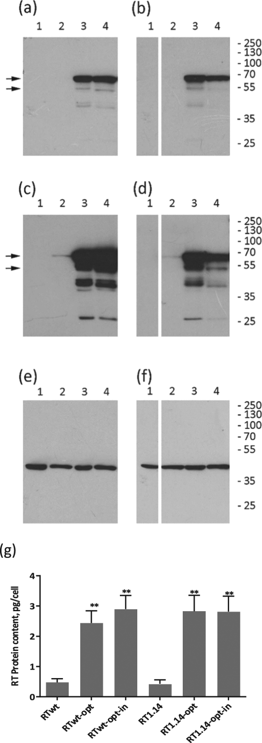
Figure 1.

Expression of RT variants in eukaryotic cells. (a–f) Western blotting of the lysates of HeLa cells transfected with vector pVax1 (lane 1), and pVax-based plasmids expressing RTwt (lane 2 a,c,e), RTwt-opt (lane 3 a,c,e), RTwt-opt-in (lane 4 a,c,e), RT1.14 (lane 2 b,d,f), RT1.14-opt (lane 3 b,d,f), and RT1.14-opt-in (lane 4 b,d,f). Blots of RTwt and RT1.14 variants were processed in parallel. Blots were stained with rabbit polyclonal anti-RT antibodies59 (a–d) and then stripped and re-stained with monoclonal anti-actin antibodies. (e,f) Positions of the relevant molecular mass markers are given to the right in kDa. Arrows point at the p66 and p51 RT subunits. Panels (a,b) represent results of a 0.5 min exposure of X-ray film with a blot, while panels (c,d) represent results of a 10 min exposure. Full-length blots are presented in Supplementary Fig. 2. (g) The average amount of RT protein expressed per HeLa cell transfected with RT variant genes. **p < 0.01 (as compared to non-optimized RT variants) using the Mann–Whitney U-tests. The graph represents the results of three independent runs, each done in duplicate, and the error bars represent the SD.
