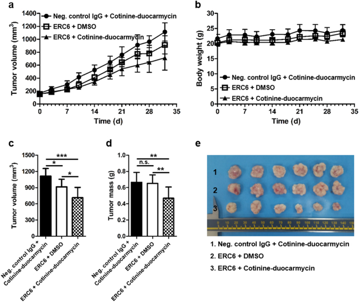
Fig. 6. Efficacy of ERC6-complexed cotinine-duocarmycin in a mouse xenograft tumor model.
a The A549 cells were subcutaneously injected into the left and right flanks of Balb/c-nude mice. When the tumor volume reached 150 mm3, the mice were randomly divided into three groups (n = 4/group) and treated for 5 weeks. Each group was intraperitoneally injected with ERC6-complexed cotinine-duocarmycin (▲), ERC6 and DMSO (□) or negative control IgG and cotinine-duocarmycin (●), and the tumor volumes were measured for 32 days. b Body weights were monitored during the treatment period. c The average tumor volume on day 32. The results are shown as the mean ± SD; *P < 0.05, **P < 0.01 compared with the group; Student’s t test. d The average dissected tumor mass at sacrifice. e Tumor tissues from three treatment groups on day 32

