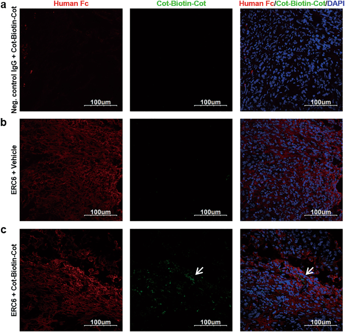
Fig. 7. Tissue distribution of ERC6 and Cot-Biotin-Cot peptide in a mouse xenograft tumor model.
Representative images from confocal microscopy. The A549 cells were subcutaneously injected into the left flanks of Balb/c-nude mice. When the tumors reached 500 mm3, the mice were intraperitoneally injected with a negative control IgG + Cot-Biotin-Cot peptide, b ERC6 + vehicle, or (c) ERC6-complexed Cot-Biotin-Cot peptide. The tumors were dissected at 24 h post-injection, and the tumor sections were stained with an Alexa 594-labeled anti-human Fc (red) and Alexa 488-labeled streptavidin (green). The nuclei were stained with 4′,6-diamidino-2-phenylindole (DAPI; blue). Image magnification, ×40

