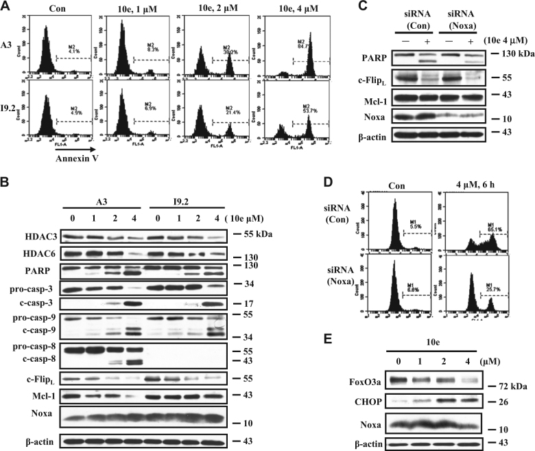
Fig. 4. Noxa plays a more important role than c-Flip and Mcl-1 in the apoptosis induction by 10e treatment.
A, B A3 cells expressing caspase-8 and I9.2 cells detecting caspase-8 were treated with 10e at the indicated concentrations for 6 h. Apoptotic cells were determined using FACS analysis after staining with Annexin V (A). The relative levels of each protein were measured using specific antibodies with Western blot analysis (B). C, D I9.2 cells were transfected with Noxa siRNA for 16 h, then treated with 4 μM 10e for additional 6 h. The levels of PARP, c-FlipL, Mcl-1, Noxa, and β-actin were determined by Western blotting (C). The apoptotic cells were quantified using FACS after staining with Annexin V-FITC (D). E I9.2 cells were treated with 10e at the indicated concentration for 6 h, the levels of FoxO3a, CHOP, Noxa, and β-actin were measured by western blot analysis

