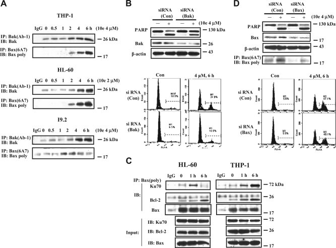
Fig. 5. Bak and Bax are activated in 10e-treated cells and contribute to the apoptosis induction.
A The activated Bak or Bax proteins in THP-1, HL-60, and I9.2 cells treated with 2 μM or 4 μM 10e for the given times were immunoprecipitated with the anti-Bak(Ab-1) or anti-Bax (6A7) antibody (detecting the active forms), respectively, followed by the western blotting using poly anti-Bak or anti-Bax antibody. B THP-1 cells were transfected with Bak siRNA or a negative control siRNA for 30 h, then treated with 4 μM 10e for additional 6 h. The levels of PARP and Bak were determined by western blotting. The apoptotic cells were measured by FACS after staining with Annexin V-FITC. C The THP-1 and HL-60 cell lysates treated with 4 μM or 2 μM 10e for 6 h were immunoprecipitated with anti-Bax antibody and immunoblotted with an anti-Ku70, Bax, or Bcl-2 antibody. D THP-1 cells were transfected with Bax siRNA or a negative control siRNA for 30 h, then treated with 4 μM 10e for additional 6 h. The levels of PARP and Bax were determined by western blotting. The active form of Bax was detected with IP. The apoptotic cells were measured by FACS after staining with Annexin V-FITC

