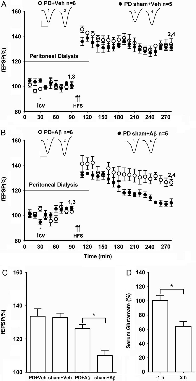
Figure 3.
Acute peritoneal dialysis prevents Aβ-mediated inhibition of LTP. (A,B) Time course graphs showing the effect of peripheral peritoneal dialysis on Aβ-mediated inhibition of LTP. (A) HFS induced robust LTP in animals that underwent peritoneal dialysis (gray bar) for 2 h, starting 30 min before i.c.v. injection of vehicle and finishing 30 min post-HFS (PD + Veh) (P < 0.05 compared with pre-HFS baseline). Similarly, in animals that underwent the same surgical protocol, but without initiating peritoneal dialysis (PD sham + Veh), HFS also triggered stable LTP (P < 0.05 compared with pre-HFS baseline). (B) Whereas, the surgical protocol had no effect on Aβ-mediated inhibition of LTP (PD sham + Aβ) (P > 0.05 compared with pre-HFS baseline) HFS induced robust LTP in animals that had 2 h peritoneal dialysis with Aβ (PD + Aβ) (P < 0.05 compared with pre-HFS baseline). Insets show representative fEPSP traces at the times indicated. Calibration bars: vertical, 1 mV; horizontal, 10 ms. (C) Summary bar chart comparing the magnitude of synaptic potentiation at 3 h post-HFS between treatment groups. Values are the mean ± SEM fEPSP amplitude expressed as a percentage of the pre-HFS baseline. (D) Compared with pretreatment values peritoneal dialysis significantly reduced serum concentration of glutamate 2 h after commencing treatment (n = 4, paired t-test). *P < 0.05.

