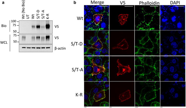
Figure 6. Localization of IFNGR1 mutant proteins.

(a) HEK cells were transfected with plasmids encoding V5-tagged IFNGR1 Wt, K-R, or the S/T-A, S/T-D mutants for 48 h. Cells were then treated with membrane impermeable biotin, harvested, and probed for IFNGR1 to determine plasma membrane localization. Wt (No Bio) = transfected, no biotin control, UT, untransfected. n = 2 independent experiments. (b) A549 cells were transfected with plasmids encoding V5-tagged IFNGR1 Wt, K-R, or the S/T-A, S/T-D mutants on coverslips for 48 h. Cells were then fixed, permeabilized and stained for DAPI for nuclear staining, phalloidin to visualize the plasma membrane, and V5 to examine IFNGR. Scale bar = 25 μm.
