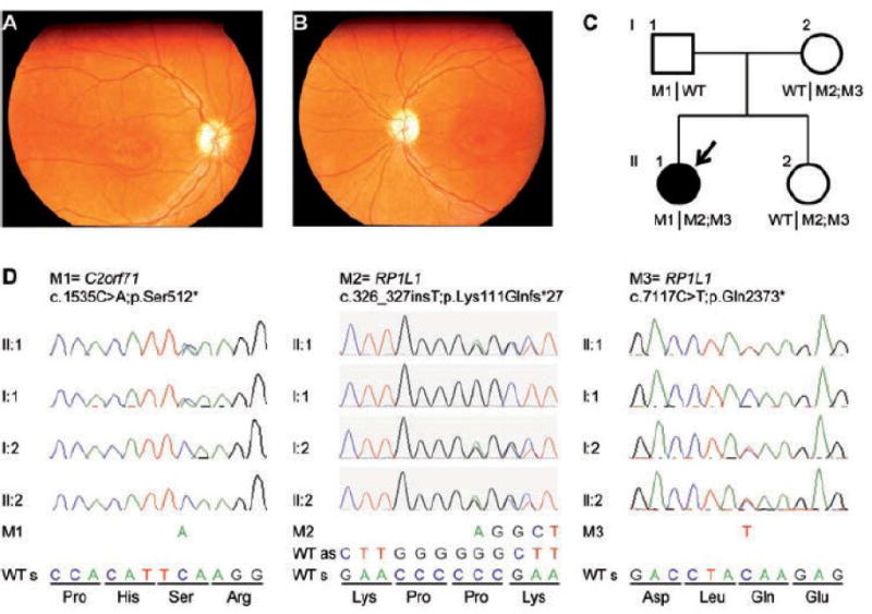
Fig. 1. Clinical and genetic characterization of a family with syndromic retinal dystrophy.

(A, B) Fundus photographs (A = right eye, B = left eye) taken at 18 years of age show normal retinal vessels, maculae with small wall-shaped degenerative changes and pale optic discs in both eyes. (C) Pedigree of the family. The arrow indicates the person in whom exome sequencing was performed. (D) Validation by Sanger sequencing of the identified RP1L1 c.[326_327insT;7117C>T] and C2orf71 c.1535C>A mutations in the affected individual, the unaffected sibling and her parents. Segregation analyses by Sanger sequencing point towards a digenic inheritance. As = antisense, M = mutation, s = sense, WT = wild-type.
