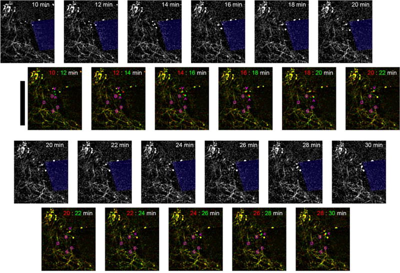
Figure 10. Dynamic neurite sprouting following microelectrode insertions in V1 in Thy1-YFP mice.
In vivo multiphoton microscopy of YFP labeled neurites show growth cones near the microelectrode minutes after insertion. Overlay images show before (red) and after (green) for each pair of time points. Yellow indicates stationary features. Pink circles identify ‘elbows’ of neurites to show there is no bulk tissue movement around the implant. Pink arrowheads point to growth cones moving towards the implant. Depth of imaging is between 0–50 µm. Scalebar = 100 µm.

