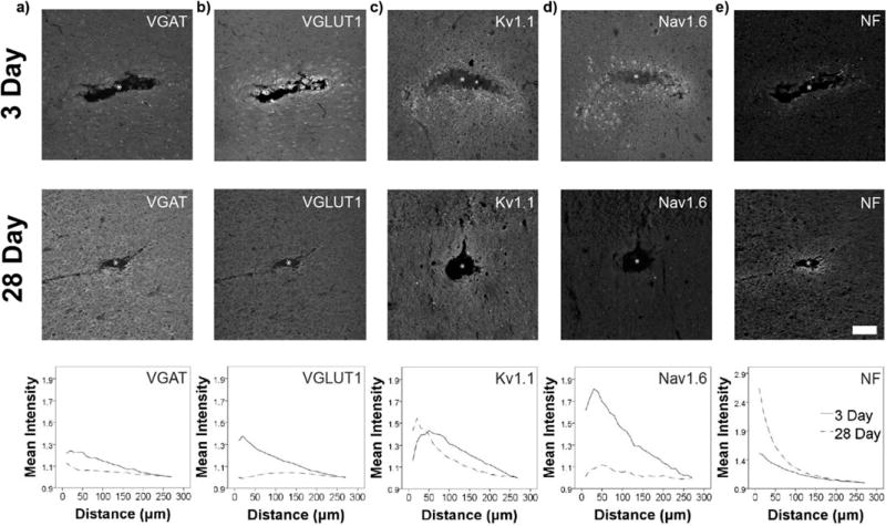
Figure 9. Evidence for plasticity in markers of excitability surrounding devices.
Immunohistochemistry at 3 days and 28 days post single shank silicon array implantation in rat primary motor cortex (n=11). a,b) At 3 days, VGLUT1 and VGAT are both significantly elevated (**p≤0.001) and VGLUT1 intensity is significantly greater than VGAT (**p≤0.001, within the first 40µm). By four weeks, VGAT intensity is significantly elevated (*p≤0.05) and is greater than VGLUT1 (*p≤0.05) in the first 40µm of the device. c,d) Early observations suggest that Nav1.6 expression is initially upregulated at the device interface, while Kv1.1 expression is increased surrounding the device by the four week time point (statistical analysis omitted due to limited sample size). e) NF is significantly elevated nearest the implant site based on quantitative immunohistochemistry (as previously reported42), suggesting possible axonal sprouting. White asterisk (*) – a,b,e) Adapted with permission from42. Copyright 2017, The American Physiological Society.

