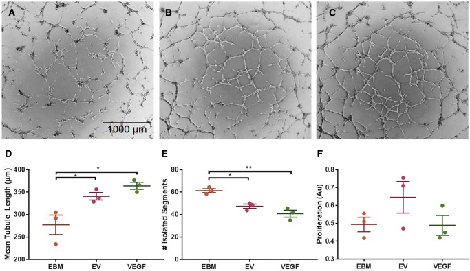
Figure 3.
Tubule formation assay shows pro-angiogenic effect of EVs after 11 h. (A) HUVECs incubated in EBM alone, (B) with EVs, and (C) with 1 ng/ml VEGF. (D) Quantitative analysis of mean tubule length for each group. (E) Quantitative analysis of isolated segment, indicating disorganized tubule formation. (F) BrdU proliferation assay shows no significant difference in HUVEC proliferation after incubation with EBM alone, EVs, or VEGF (n = 3, *P < 0.05, **P < 0.01).

