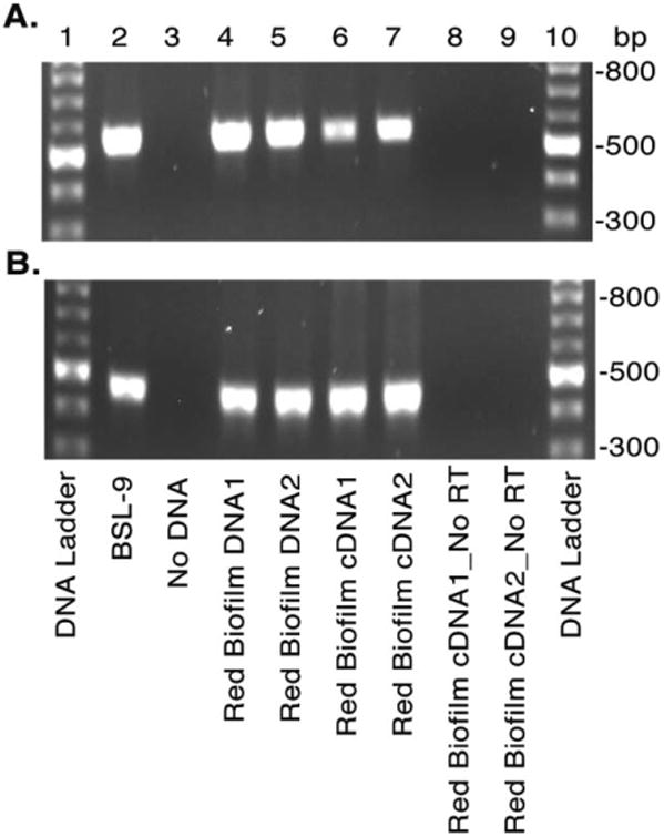
Fig. 6.

Evidence for environmental photoarsenotrophy activity determined by arxA gene expression in situ. Red biofilm-like microbial mats within hot springs of Paoha Island Mono Lake, CA were collected and the RNA and DNA extracts analysed for arxA (arxA_1824_deg_F and arxA_2380_deg_R primers) (A) and 16S rRNA (B) by PCR and RT-PCR respectively. Agarose gel electrophoresis of the PCRs and RT-PCRs are shown in A and B. For both panels lanes correspond to: DNA ladder (1, 10), BSL-9 genomic DNA (2), water negative control (3), environmental DNA (4–5), and cDNA of environmental RNA samples with (6–7) and without (8–9) reverse transcriptase.
