
Fig. 3.
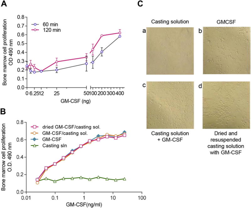
GM-CSF retains proliferative capacity in bone marrow cells following MN patch fabrication and dissolution. (A) Dose-responsive cellular activation of bone marrow cells by murine GM-CSF in PBS at 60 and 120 min. (B) Bone marrow cells (5 × 10⁵ cells/well) from naïve mice were incubated for 48 h with (a) “first-cast” casting solution, (b) GM-CSF dissolved in PBS, (c) GM-CSF mixed 1:1 with casting solution, or (d) GM-CSF in casting solution air-dried in microcentrifuge tubes and reconstituted in PBS. Bioactivity was measured by determining cell proliferation at 48 h post-exposure. (C) Bone marrow cells grown in tissue culture were imaged at 48 h post-exposure to GM-CSF in formulations described in (B).