
Figure 3.
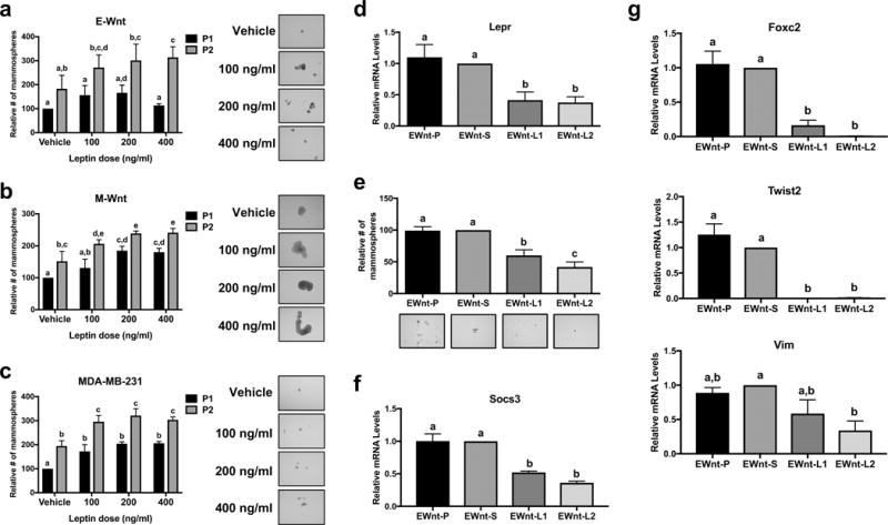
Leptin stimulates mammosphere formation in triple-negative mammary tumor cells. Mammosphere formation in (a) E-Wnt, (b) M-Wnt, and (c) MDA-MB-231 cells was assessed at the end of propagation 1 (P1), during which the cells were treated for 7 days with leptin, and following propagation 2 (P2), in which the spheres from P1 were dissociated and then replated with the same treatments for another 7 days. Representative images of mammospheres at the end of P2 are shown at x10 magnification. (d) Lepr expression in parental E-Wnt cells (EWnt-P) as well as E-Wnt cells stably transfected with a scrambled shRNA plasmid (EWnt-S) or shRNA to Lepr (EWnt-L1 and EWnt-L2) was measured by quantitative RT-PCR. (e) Mammosphere formation was assessed in EWnt-P, EWnt-S, EWnt-L1, and EWnt-L2 cells after a 7-day incubation in mammosphere media. Socs3 (f) and Foxc2, Twist2, and Vim (g) gene expression was measured by quantitative RT-PCR. Different letters indicate significant differences, P<0.05.