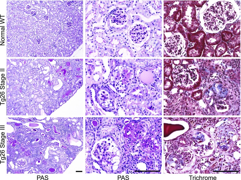
Figure 1.
Stages of progressive kidney disease pathology in Tg26 mice. Photomicrographs of representative kidney sections from normal mice (top row) and mice with stage 2 (middle row) and stage 3 (bottom row) disease with periodic acid–Schiff (PAS; left and middle columns) and trichrome stains (right column). Kidney cortex of normal mice is well preserved; glomeruli show open capillary loops, and no significant mesangial expansion, epithelial or inflammatory cell proliferation (PAS), or significant collagen is seen in the glomeruli (trichrome). Kidney cortex of stage 2 mice shows focal tubulointerstitial changes, including signs of focal tubular injury, luminal distension and epithelial flattening, and focal tubular microcystic change with prominent hyaline cast formation (PAS). Glomeruli of stage 2 mice show focal global or segmental collapsing lesions characterized by collapse of the tuft with associated epithelial changes, including proliferation within Bowman’s space, protein reabsorption, and vacuolization (PAS). Trichrome staining of stage 2 glomeruli shows focal segmental collagen deposition in the tuft largely confined to lesional glomeruli. Kidney cortex of stage 3 mice exhibits widespread chronic changes with extensive tubular microcystic change and few viable tubules noted. In stage 3 mice, many sclerosed glomeruli are present along with glomeruli showing changes similar to those seen in stage 2 mice, and trichrome stain shows segmental to global collagen deposition in the tuft of most glomeruli. Magnification, ×100 in the left column; ×400 in the middle and right columns. Scale bar, 100 μm.

