Figure 1.
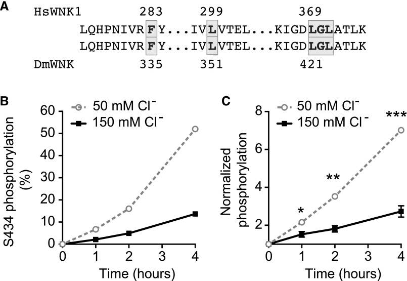
D. melanogaster With No Lysine kinase (DmWNK) autophosphorylation is inhibited by chloride in vitro. (A) Alignment of the chloride binding domain of human With No Lysine kinase 1 (HsWNK1) and DmWNK kinase domains shows conservation of this region. Chloride binding residues7 are highlighted. (B) DmWNK kinase domain (amino acids 261–534) was purified (Supplemental Figure 1A) and allowed to autophosphorylate in vitro in buffers containing 50 or 150 mM NaCl. Autophosphorylation at Ser 434 (Supplemental Figure 1B) was monitored at 0, 1, 2, and 4 hours using mass spectrometry. (C) DmWNK kinase domain autophosphorylation was performed as in B. The reaction was terminated, protein was electrophoresed using SDS-PAGE, and the Pro-Q Diamond phosphoprotein stain was used to determine kinase autophosphorylation followed by band densitometry for quantification. Three independent experiments were performed (Supplemental Figure 1C). Results were normalized to the 0-hour time point for each experiment. In this figure and subsequent figures, mean±SEM is shown; 50 versus 150 mM chloride values at each time point were compared using multiple t tests, with Holm–Sidak correction for multiple comparisons. Adjusted P values are shown. *P=0.03 at 1 hour; ***P=0.003 at 2 hours; ***P<0.001 at 4 hours.

